
Hydrocele is a collection of fluid in the thin sac that surrounds a testicle, which makes one side of the scrotum swell like a small water balloon. In adults it is usually painless and harmless, but it rarely disappears on its own, and an ultrasound is often used to confirm the diagnosis and check the testicle behind the fluid. Treatment is only needed when the swelling is large, uncomfortable or the diagnosis is uncertain, and surgery (hydrocelectomy) is the standard cure.
Sources: MedlinePlus (NIH) — encyclopedia entry; Cleveland Clinic — Causes, Symptoms, Diagnosis & Treatment
- A hydrocele is fluid trapped between the two layers of the tunica vaginalis, the lining around the testicle; about 1% of adult men have one.
- In adults it can be idiopathic (no clear cause) or secondary to infection, injury, surgery, a hernia or, rarely, a testicular tumor, so the testicle itself must always be checked.
- Scrotal ultrasound is the main imaging test: it confirms fluid, shows whether the testicle behind it is normal and separates the fluid from a hernia, cyst or varicocele.
- A small swelling that causes no symptoms can simply be watched. Surgery is the standard treatment; needle drainage alone lets the fluid come back quickly, and sclerotherapy carries a higher recurrence risk.
- The condition usually does not cause infertility, but any procedure near the epididymis and vas deferens should be planned carefully in men who still want children.
- What is a hydrocele?
- Hydrocele causes in adults
- Hydrocele symptoms
- How a hydrocele is diagnosed: exam and ultrasound
- When should an adult hydrocele be treated?
- Hydrocele treatment: hydrocelectomy vs aspiration and sclerotherapy
- Recovery after hydrocelectomy
- Does a hydrocele affect fertility?
- When to see a doctor
- FAQs about hydrocele
What is a hydrocele?
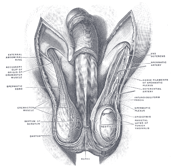
Each testicle sits inside a double-layered lining called the tunica vaginalis. Normally only 1–2 mL of lubricating fluid lies between these layers. The swelling forms when more fluid collects there than the lining can reabsorb, either because too much is produced or because drainage is poor.
Sources: Ultrasound International Open (PMC) — Patil et al. 2015 — Fluid within the scrotal spaces; MedlinePlus (NIH) — encyclopedia entry
Doctors divide hydroceles into two types. In a communicating hydrocele, a channel left over from fetal development still connects the scrotum to the abdomen, so fluid can move back and forth and the swelling may change size during the day. In a non-communicating hydrocele, that channel has closed and the fluid stays around the testicle; the size usually stays the same or grows very slowly. Most adult cases are non-communicating, and either type can affect one or both sides.
Sources: Cleveland Clinic — Causes, Symptoms, Diagnosis & Treatment; Patient.info (professional) — clinical overview
Hydroceles are much more common in newborn boys, where about 10% are affected and most clear up within the first year. About 1% of adult men have one, although researchers note that the true adult prevalence is not well studied. It is one of the leading causes of painless scrotal swelling, and in adults it is more common in older men.
Sources: Cleveland Clinic — Causes, Symptoms, Diagnosis & Treatment; Cochrane Review (PMC) — Shakiba et al. 2014 — Aspiration and sclerotherapy versus hydrocoelectomy for treating hydrocoeles
Hydrocele causes in adults
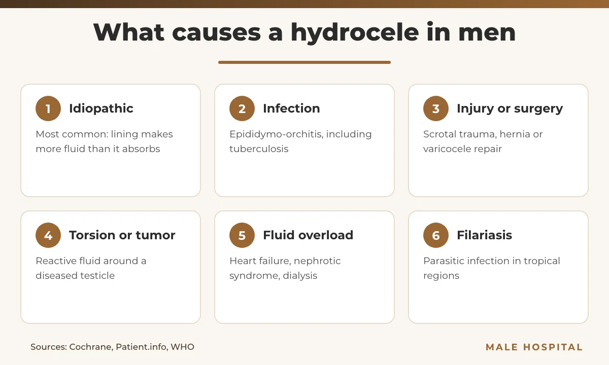
Most acquired hydroceles in men are idiopathic, meaning no single trigger is found; the lining simply makes more fluid than it absorbs. This is called a primary form. A secondary one, by contrast, is a reaction to another problem in the scrotum or body, and finding that problem matters more than the fluid itself.
Sources: Cochrane Review (PMC) — Shakiba et al. 2014 — Aspiration and sclerotherapy versus hydrocoelectomy for treating hydrocoeles; Ultrasound International Open (PMC) — Patil et al. 2015 — Fluid within the scrotal spaces
Secondary causes
- Inflammation or infection of the testicle or epididymis (epididymo-orchitis), including tuberculosis.
- Injury to the scrotum or testicle.
- Testicular torsion, a twisted testicle.
- Previous scrotal or groin surgery, such as hernia repair or treatment of a varicocele.
- Testicular tumor, which can produce a reactive hydrocele.
- Generalized fluid retention, for example from heart failure or nephrotic syndrome, after kidney transplantation or after radiation therapy.
Sources: Patient.info (professional) — clinical overview; Cochrane Review (PMC) — Shakiba et al. 2014 — Aspiration and sclerotherapy versus hydrocoelectomy for treating hydrocoeles
A communicating hydrocele can occasionally appear for the first time in adulthood when pressure inside the abdomen rises, for example in men on peritoneal dialysis or with fluid overload. In this situation an inguinal hernia may sit alongside the fluid, because both use the same open channel.
Sources: Patient.info (professional) — clinical overview; MedlinePlus (NIH) — encyclopedia entry
Filarial hydrocele
In tropical regions, an important cause of hydrocele is lymphatic filariasis, a parasitic infection spread by mosquitoes in tropical regions. The World Health Organization’s baseline estimate was 25 million men with this complication, and WHO notes that surgery can relieve most of these cases. Men who have lived in endemic areas should mention this to their doctor.
Source: WHO — Lymphatic filariasis fact sheet
Hydrocele symptoms
The main sign is a smooth, round or oval swelling of the scrotum, on one or both sides, that feels like a water balloon. It is usually painless, and MedlinePlus notes that the right side is affected more often. Because the testicle sits behind the fluid, it may be hard to feel through a large hydrocele.
Source: MedlinePlus (NIH) — encyclopedia entry
Other symptoms come from size and weight rather than from damage to the testicle:
- a feeling of heaviness or dragging in the scrotum,
- discomfort or pain, especially as the swelling grows,
- swelling that grows through the day and shrinks overnight (typical of a communicating hydrocele),
Source: Cleveland Clinic — Causes, Symptoms, Diagnosis & Treatment
Pain, tenderness, redness, fever or a hard lump in the testicle are not typical of simple fluid around the testicle. They suggest an underlying problem such as infection, torsion or a tumor and need prompt medical review, as described in the red flags section below.
Sources: Patient.info (professional) — clinical overview; MedlinePlus (NIH) — encyclopedia entry
How a hydrocele is diagnosed: exam and ultrasound
Diagnosis starts with a history and examination. The doctor checks whether the swelling is confined to the scrotum or extends into the groin, presses on the groin and asks you to cough to look for a hernia, and shines a light through the scrotum (transillumination). Clear fluid lets light pass, while solid tissue does not. If the swelling changes size when pressure is applied, an inguinal hernia is more likely.
Sources: Cleveland Clinic — Causes, Symptoms, Diagnosis & Treatment; MedlinePlus (NIH) — encyclopedia entry
Transillumination is helpful but not decisive. The fluid can hide a more urgent problem, so a glowing scrotum does not exclude a tumor, infection or torsion underneath.
Source: Patient.info (professional) — clinical overview
Why scrotal ultrasound matters
Ultrasound is the most common imaging test for this condition and the key test in adults. It uses a high-frequency probe, usually above 10 MHz, to scan both testicles in at least two planes. Simple fluid appears as dark (anechoic) fluid around a normal testicle; thin dividing walls (septa), small stones or scattered echoes from protein or cholesterol can appear in long-standing cases.
Sources: Cleveland Clinic — Causes, Symptoms, Diagnosis & Treatment; Ultrasound International Open (PMC) — Patil et al. 2015 — Fluid within the scrotal spaces
The most important job of the scan is to look at the testicle and epididymis behind the fluid. Ultrasound separates lesions inside the testicle from those outside it, and Doppler shows blood flow, which helps distinguish the fluid from a varicocele or torsion. It is especially indicated when the testicle cannot be felt clearly, when there is tenderness, or when the scan shows internal echoes. German andrology guidance states that treatment decisions should be based on high-resolution ultrasound.
Sources: Ultrasound International Open (PMC) — Patil et al. 2015 — Fluid within the scrotal spaces; Patient.info (professional) — clinical overview; Der Urologe (PubMed) — Kliesch 2014 — management of complications
Other tests
If a testicular tumor is suspected, blood tests for tumor markers such as alpha-fetoprotein (AFP) and hCG are used, and diagnostic needle aspiration should be avoided because it could spread malignant cells. A CT scan is occasionally used when the picture is unclear.
Sources: Patient.info (professional) — clinical overview; Cleveland Clinic — Causes, Symptoms, Diagnosis & Treatment
When should an adult hydrocele be treated?
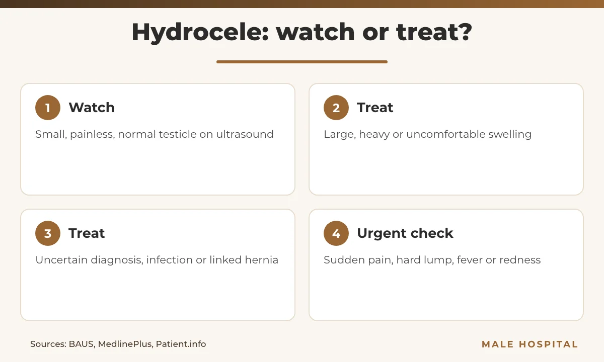
Many adult hydroceles never need treatment. If the swelling is small and not bothersome, observation is a reasonable choice once the ultrasound has shown a normal testicle. Unlike in babies, though, adult hydroceles rarely resolve on their own, and there are no medicines that shrink one.
Sources: BAUS — patient leaflet A24/127 (2024); MedlinePlus (NIH) — encyclopedia entry; Cleveland Clinic — Causes, Symptoms, Diagnosis & Treatment
Treatment is usually considered when:
- the hydrocele is large, heavy or uncomfortable, or interferes with daily activity or sex,
- the diagnosis is uncertain or the testicle cannot be assessed,
- the hydrocele becomes infected,
- it comes back after needle drainage,
- it is linked to an inguinal hernia, which should be repaired promptly.
Sources: Patient.info (professional) — clinical overview; MedlinePlus (NIH) — encyclopedia entry
The decision is about symptoms, not size alone. Asymptomatic findings do not need correction, and in younger men the advantages and disadvantages of an operation, including possible effects on fertility, should be discussed in detail first.
Source: Der Urologe (PubMed) — Kliesch 2014 — management of complications
Hydrocele treatment: hydrocelectomy vs aspiration and sclerotherapy
Hydrocelectomy (surgery)
Open hydrocelectomy is considered the standard treatment in adults. It is normally done under general or spinal anesthesia; local anesthesia is possible but unusual. Through a small scrotal incision the surgeon drains the fluid and then either “bunches up” the sac with absorbable stitches (plication, often called Lord’s procedure) or turns it inside out and closes it behind the testicle (eversion, often called Jaboulay’s procedure). A very thick sac may be removed completely. The skin is closed with dissolvable stitches.
Sources: Cochrane Review (PMC) — Shakiba et al. 2014 — Aspiration and sclerotherapy versus hydrocoelectomy for treating hydrocoeles; BAUS — patient leaflet A24/127 (2024); Urology (PubMed) — Tsai et al. 2019 — Recurrence and complications of 3 techniques
A groin (inguinal) approach is preferred for a communicating hydrocele, because the open channel to the abdomen must be closed, and it is essential when a tumor is suspected.
Sources: Patient.info (professional) — clinical overview; Cleveland Clinic — Causes, Symptoms, Diagnosis & Treatment
How do the techniques compare? In a US series of 276 operations for idiopathic hydrocele, about 6% needed repeat surgery for recurrence, with no difference between Jaboulay’s repair, excision and Lord’s repair. Complications such as hematoma, testicular pain and infection occurred in 11.6%, and Lord’s repair had the lowest overall complication and hematoma rates. The British Association of Urological Surgeons (BAUS) quotes recurrence after surgery at between 1 in 50 and 1 in 250 patients.
Sources: Urology (PubMed) — Tsai et al. 2019 — Recurrence and complications of 3 techniques; BAUS — patient leaflet A24/127 (2024)
Needle aspiration
Aspiration means drawing the fluid out with a needle. It gives quick relief, but the fluid usually comes back, often quickly, and BAUS describes it as neither curative nor standard practice; it can also introduce infection. Aspiration alone is mainly used for temporary relief in older men who are not fit for surgery.
Sources: BAUS — patient leaflet A24/127 (2024); MedlinePlus (NIH) — encyclopedia entry; Patient.info (professional) — clinical overview
Aspiration with sclerotherapy
Sclerotherapy adds a step: after draining the fluid, a sclerosing agent is injected to make the two layers of the sac stick together. Agents used in trials include sodium tetradecyl sulfate, tetracycline or doxycycline, phenol and polidocanol. It avoids an incision, but it can cause pain and, because the chemical can inflame the epididymis, epididymal obstruction.
Sources: Cochrane Review (PMC) — Shakiba et al. 2014 — Aspiration and sclerotherapy versus hydrocoelectomy for treating hydrocoeles; Patient.info (professional) — clinical overview
A Cochrane review of four small randomized trials (275 men) found that recurrence was significantly higher after sclerotherapy than after surgery, while short-term cure rates were not clearly different. Sclerotherapy was cheaper and meant about six fewer days off work in one trial. A 2023 meta-analysis of five small trials (335 men) reached the same conclusion and recommended sclerotherapy mainly for men at high surgical risk or those who wish to avoid surgery. Both reviews rated the evidence as low quality.
Sources: Cochrane Review (PMC) — Shakiba et al. 2014 — Aspiration and sclerotherapy versus hydrocoelectomy for treating hydrocoeles; Surgical Endoscopy (PubMed) — Shakiba et al. 2023 — systematic review and meta-analyses
| Option | How it works | Recurrence | Best suited for |
|---|---|---|---|
| Observation | No procedure; review if symptoms change | Swelling persists | Small, painless hydrocele with a normal testicle on ultrasound |
| Hydrocelectomy (Lord’s plication or Jaboulay eversion) | Fluid drained; sac folded, everted or removed | Lowest; about 1 in 50 to 1 in 250 (BAUS), 6% in one large series | Most symptomatic men, including younger men and suspected tumor (inguinal approach) |
| Aspiration alone | Fluid removed with a needle | High; fluid usually returns quickly | Short-term relief in older men unfit for surgery |
| Aspiration + sclerotherapy | Fluid removed, sclerosant injected | Higher than surgery; may need repeat sessions | Men at high surgical risk when fertility is not a concern |
Sources: BAUS — patient leaflet A24/127 (2024); Urology (PubMed) — Tsai et al. 2019 — Recurrence and complications of 3 techniques; Cochrane Review (PMC) — Shakiba et al. 2014 — Aspiration and sclerotherapy versus hydrocoelectomy for treating hydrocoeles; Patient.info (professional) — clinical overview
Recovery after hydrocelectomy
Most men return to normal light activities within a few days and need about a week off work, longer for physically demanding jobs, while full healing takes several weeks.
Source: Cleveland Clinic — Causes, Symptoms, Diagnosis & Treatment
BAUS gives practical aftercare advice:
- Expect swelling and bruising of the scrotum for several days to a few weeks; almost every patient has some.
- Wear a scrotal support, tight supportive underwear or cycling shorts for comfort.
- Simple painkillers such as paracetamol or ibuprofen and wrapped ice packs (never directly on the skin) help in the first few days.
- The dissolvable stitches usually disappear within two to three weeks.
- Avoid heavy lifting and strenuous exercise for at least four weeks.
- Contact your surgical team if bruising, swelling or pain gets worse day by day.
Source: BAUS — patient leaflet A24/127 (2024)
After surgery the treated testicle almost always feels “bulkier” than the other side, because the folded sac remains. This is expected and does not mean the fluid has returned. A routine follow-up visit is not always needed, but any new swelling after several months should be checked.
Source: BAUS — patient leaflet A24/127 (2024)
Possible complications
According to BAUS, a blood collection (hematoma) and wound or testicular infection each occur in between 1 in 10 and 1 in 50 patients. Recurrence, injury to the epididymis, vas deferens or testicular blood supply, and chronic scrotal pain each occur in between 1 in 50 and 1 in 250. Smoking raises the risk of complications, so stopping before surgery is advised.
Source: BAUS — patient leaflet A24/127 (2024)
Does a hydrocele affect fertility?
In most men, no. A hydrocele surrounds the testicle but does not usually damage sperm production, and Cleveland Clinic states that hydroceles usually don’t cause infertility. A very large, tense collection can compress the testicle and increase resistance to its blood flow on Doppler ultrasound; draining the fluid restored normal flow in reported cases.
Sources: Cleveland Clinic — Causes, Symptoms, Diagnosis & Treatment; Ultrasound International Open (PMC) — Patil et al. 2015 — Fluid within the scrotal spaces
The main fertility concern is the treatment, not the fluid itself. The epididymis and vas deferens lie right next to the sac, and BAUS quotes accidental injury to these structures in between 1 in 50 and 1 in 250 operations. Infertility from surgery is possible mainly if the vas is injured on both sides. Sclerotherapy can inflame and block the epididymis, which is why it is usually reserved for men for whom fertility is not a concern.
Sources: BAUS — patient leaflet A24/127 (2024); Patient.info (professional) — clinical overview
For younger men who still want children, German andrology guidance recommends discussing the wish for children before surgery and, where appropriate, offering sperm freezing (cryopreservation) beforehand. None of the randomized trials comparing sclerotherapy with surgery measured sperm quality, so there is no direct trial evidence on fertility after either method. If you are already trying to conceive, a semen analysis before treatment gives a useful baseline, and our guide to male infertility explains other causes worth checking.
Sources: Der Urologe (PubMed) — Kliesch 2014 — management of complications; Cochrane Review (PMC) — Shakiba et al. 2014 — Aspiration and sclerotherapy versus hydrocoelectomy for treating hydrocoeles
When to see a doctor
Any new scrotal swelling should be examined by a doctor, even if it is painless, because other causes of a lump in the scrotum, including testicular cancer, must be ruled out. Book a routine urology appointment if you notice a soft, painless swelling that is getting bigger, feels heavy or bothers you.
Sources: MedlinePlus (NIH) — encyclopedia entry; Cleveland Clinic — Causes, Symptoms, Diagnosis & Treatment
Seek urgent or emergency care if you have:
- sudden or severe pain in the testicle or scrotum, especially with swelling (possible torsion, which can cost the testicle if not treated quickly),
- a hard lump or a testicle that feels different, or a testicle that cannot be felt through the swelling,
- redness, warmth, tenderness or fever (possible infection),
- a painful bulge in the groin (possible inguinal hernia),
- after surgery: swelling, bruising or pain that worsens day by day, discharge from the wound or a high temperature.
Sources: MedlinePlus (NIH) — encyclopedia entry; Patient.info (professional) — clinical overview; BAUS — patient leaflet A24/127 (2024)
If you have a scrotal swelling and would like an expert opinion, you can arrange a confidential consultation with our urologist-andrologist, Assoc. Prof. Dr. Zülfü Sertkaya, via WhatsApp. The right option and its cost depend on the examination and ultrasound findings.
FAQs about hydrocele
Can a hydrocele go away on its own in adults?
Rarely. Hydroceles in babies often disappear within the first year, but in adults they seldom resolve without treatment. Fluid that follows an injury or infection may settle once the underlying problem heals. If it is small and painless, watching it is safe once a doctor has examined you and an ultrasound has shown a healthy testicle.
Sources: MedlinePlus (NIH) — encyclopedia entry; Cleveland Clinic — Causes, Symptoms, Diagnosis & Treatment
Is a hydrocele dangerous?
A simple hydrocele is benign and does not turn into cancer. The risk lies in what it can hide: infection, torsion or, occasionally, a testicular tumor can cause secondary fluid. That is why a new swelling needs an examination and usually an ultrasound. A communicating hydrocele can rarely be linked to an inguinal hernia, which needs repair.
Sources: Patient.info (professional) — clinical overview; Cleveland Clinic — Causes, Symptoms, Diagnosis & Treatment
Can a hydrocele be drained with a needle?
Yes, but drainage alone is a temporary fix. The fluid typically re-accumulates quickly, so it is not regarded as a cure, and every needle passage carries a small risk of infection. It is mostly used for older men who cannot have an operation. Needle drainage should not be used to diagnose a swelling when a tumor has not been excluded.
Sources: BAUS — patient leaflet A24/127 (2024); Patient.info (professional) — clinical overview
Is sclerotherapy better than surgery for a hydrocele?
Not for most men. Trials show that sclerotherapy is cheaper and allows a faster return to work, but the fluid comes back significantly more often than after hydrocelectomy. The evidence comes from small, low-quality studies. Sclerotherapy is a reasonable option for men at high surgical risk who are not planning to have children.
Sources: Cochrane Review (PMC) — Shakiba et al. 2014 — Aspiration and sclerotherapy versus hydrocoelectomy for treating hydrocoeles; Surgical Endoscopy (PubMed) — Shakiba et al. 2023 — systematic review and meta-analyses
How long does it take to recover from hydrocele surgery?
Most men go back to light activities within a few days and to desk work after about a week. Swelling and bruising can last a few weeks, and full healing takes several weeks. Heavy lifting and strenuous exercise should be avoided for at least four weeks. The stitches dissolve on their own within two to three weeks.
Sources: Cleveland Clinic — Causes, Symptoms, Diagnosis & Treatment; BAUS — patient leaflet A24/127 (2024)
Can a hydrocele come back after surgery?
Yes, but it is uncommon. BAUS quotes recurrence in between 1 in 50 and 1 in 250 patients, and a large US series found that about 6% needed repeat surgery, with no clear difference between the main techniques. Some early swelling after surgery is a normal reaction and usually settles within a few months, so it should not be mistaken for a recurrence.
Sources: BAUS — patient leaflet A24/127 (2024); Urology (PubMed) — Tsai et al. 2019 — Recurrence and complications of 3 techniques; Patient.info (professional) — clinical overview
What is the difference between a hydrocele, a spermatocele and a varicocele?
All three cause scrotal swelling but come from different structures. A hydrocele is fluid around the whole testicle, a spermatocele (epididymal cyst) is a fluid-filled cyst arising from the epididymis, and a varicocele is a cluster of widened veins above the testicle. Ultrasound distinguishes spermatoceles from hydroceles, and Doppler imaging separates a hydrocele from a varicocele or torsion.
Sources: Patient.info (professional) — clinical overview; Ultrasound International Open (PMC) — Patil et al. 2015 — Fluid within the scrotal spaces
Should I get an ultrasound for a hydrocele?
In adults, usually yes. A scrotal ultrasound confirms that the swelling is fluid, shows whether the testicle and epididymis behind it are normal, and separates it from a hernia, epididymal cyst or varicocele. It is particularly important if the testicle cannot be felt, if there is any tenderness, or before deciding on surgery.
Sources: Ultrasound International Open (PMC) — Patil et al. 2015 — Fluid within the scrotal spaces; Patient.info (professional) — clinical overview; Der Urologe (PubMed) — Kliesch 2014 — management of complications
Medically reviewed by Assoc. Prof. Dr. Zülfü Sertkaya, urologist and andrologist (FEBU), Istanbul. Last reviewed: November 2026.
This article is for general information and does not replace an individual consultation with a qualified doctor.
- MedlinePlus (NIH) — encyclopedia entry
- Cleveland Clinic — Causes, Symptoms, Diagnosis & Treatment
- BAUS — patient leaflet A24/127 (2024)
- Cochrane Review (PMC) — Shakiba et al. 2014 — Aspiration and sclerotherapy versus hydrocoelectomy for treating hydrocoeles
- Surgical Endoscopy (PubMed) — Shakiba et al. 2023 — systematic review and meta-analyses
- Urology (PubMed) — Tsai et al. 2019 — Recurrence and complications of 3 techniques
- Der Urologe (PubMed) — Kliesch 2014 — management of complications
- Ultrasound International Open (PMC) — Patil et al. 2015 — Fluid within the scrotal spaces
- Patient.info (professional) — clinical overview
- WHO — Lymphatic filariasis fact sheet